CAT/CT Scans
A CAT Scan or computed tomography (CT), is a medical imaging method that produces a volume of data that can be manipulated (through a process known as windowing) to demonstrate various bodily structures based on their ability to block the x-ray beam. A CAT is created by combining a series of x-ray views taken from different angles. Modern scanners allow for 3-D representations of structures.
CT Scan FAQ's
What is a CT Scan?
Computer Assisted Tomography (CAT), also known as CT (computerized tomography) is an x-ray technique that uses a special scanner to create cross-sectional images of the body and head. This produces “slices” like the slices in a loaf of bread. Our CT scanner performs spiral slices – the newest and fastest scanning technology available.
CT’s can image the internal portion of the organs and separate overlapping structures precisely. Unlike standard x-rays which take a picture of the whole structure being examined, CT has the ability to image that same structure one cross-section or “slice” at a time. This allows the internal body area being examined to be depicted in much greater detail than standard x-rays. CT is also able to provide clear imaging of both soft tissues, such as the brain, as well as dense tissue like bone.
Because a CT scan uses an ultra-thin, low dose x-ray beam, radiation exposure is minimized.
How will I prepare for my CT Scan?
Depending on the area of the body being imaged, you may be asked to drink a flavored mixture called contrast that will aid in the evaluation of your stomach and intestines.
Certain types of studies also require an IV contrast material, which will be administered through a vein (usually in your arm), once you are in the exam room.
If your exam requires an IV contrast material to highlight certain parts of your body, you may feel a warm sensation throughout your body and/or a metallic taste in your mouth once the IV is administered.
What will happen during the exam?
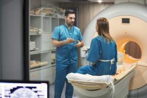
When you enter the exam room, you will be asked to lie on the CT table. The technologist will explain the procedure to you and position you on the scanning table. The table will then move to the center on the part of your body being examined. You will be able to see out both ends of the scanner, and you will be able to talk to your technologist via a two-way microphone. The table will move within the scanner during the exam. It is normal to hear whirring or clicking noises while the exam is being done.
While the exam is being done, all you need to do is relax and remain as still as possible. You may be asked to hold your breath for short periods of time.
How long is the CT scan procedure?
How long your CT procedure lasts depends on the type of scan you need. When contrast material is used, technologists may need additional time to let it move through your system. The actual scan itself can take anywhere from a few seconds to several minutes. When no contrast is used, the entire CT scan procedure generally takes about 15 to 30 minutes, which includes the time needed to prepare you.
After your imaging procedure is complete, the results are sent to your doctors within a few hours. Your doctor will then contact you to share the results of your CT scan.
Where should I go for a CT scan?
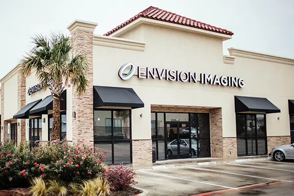
If you’re not sure where to go for a CT, Envision Radiology is an excellent choice. Included under the Envision Radiology umbrella, Envision Imaging serves patients in the Dallas-Fort Worth area of Texas, the Tulsa and Claremore areas of Oklahoma, and the Lafayette area of Louisiana. Colorado Springs Imaging serves patients in Colorado Springs, Colorado.
We recommend contacting your office of choice ahead of time to ensure that they offer CT scan imaging services, as each location is locally managed. You can also check online through the individual location pages found on our website. Just read the “Services Provided” section to see that location’s full list of offerings.
No matter which of our locations you choose, you will be treated to the same level of professionalism and compassionate care in a comfortable environment designed to put you at ease.


 CT scans are often used for emergency situations where quick action is needed, such as possible internal injuries from a car accident or other type of trauma. CT scans can be useful in many situations including:
 A CAT Scan (Computerized Axial Tomography), also referred to as a CT scan, is a computerized x-ray procedure. This provides a three-dimensional scan of the brain and other parts of the body and is used to find irregularities. This technology can be used to help guide surgeons when doing complicated surgeries, determine areas of internal damage, and to pinpoint where a disease, such as cancer, resides in your body.


