What Is an Arthrogram MRI?
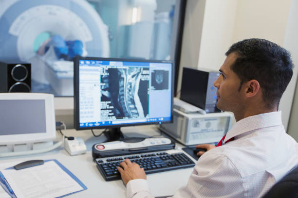
Joints are essential parts of your body that give you freedom of movement, but unfortunately, they can go wrong in many ways. An arthrogram MRI allows radiologists to pinpoint issues in your joints that standard imaging may miss. Arthrogram, also called arthrography, is a series of images taken using an X-ray, MRI, CAT scan or fluoroscopy.
Before the arthrogram MRI procedure, your joint is injected with a contrast dye, usually iodine. Using fluoroscopy, the radiologist guides the placement of the injection into the joint. The contrast dye coats the inner lining of the joint structures which makes it appear white on the arthrogram and helps highlight what’s gone wrong in your joint. Using the images, your physician can assess the function of the joint, make a diagnosis and come up with a treatment plan.


 When you arrive, you will be asked to remove all of your jewelry and any metallic objects. You may also be asked to wear medical scrubs (top and pants) or a hospital gown, or at least remove all clothing from around the joint being examined. You will then lay on a table in the examination room.


